Tendon Diagram - Anterior Cruciate Ligament Wikipedia : One peroneal tendon attaches to the outer part of the midfoot, while the other tendon runs under the foot and attaches near the inside of the arch.
byAdmin-
0
Tendon Diagram - Anterior Cruciate Ligament Wikipedia : One peroneal tendon attaches to the outer part of the midfoot, while the other tendon runs under the foot and attaches near the inside of the arch.. Raises and rotates arm in all directions. 9 photos of the foot tendons and ligaments diagram. 17 photos of the diagram of shoulder muscles and tendons. These structures work together to support the body, enable a range of movements, and send messages from the brain to. You can see a diagram of the achilles tendon below.
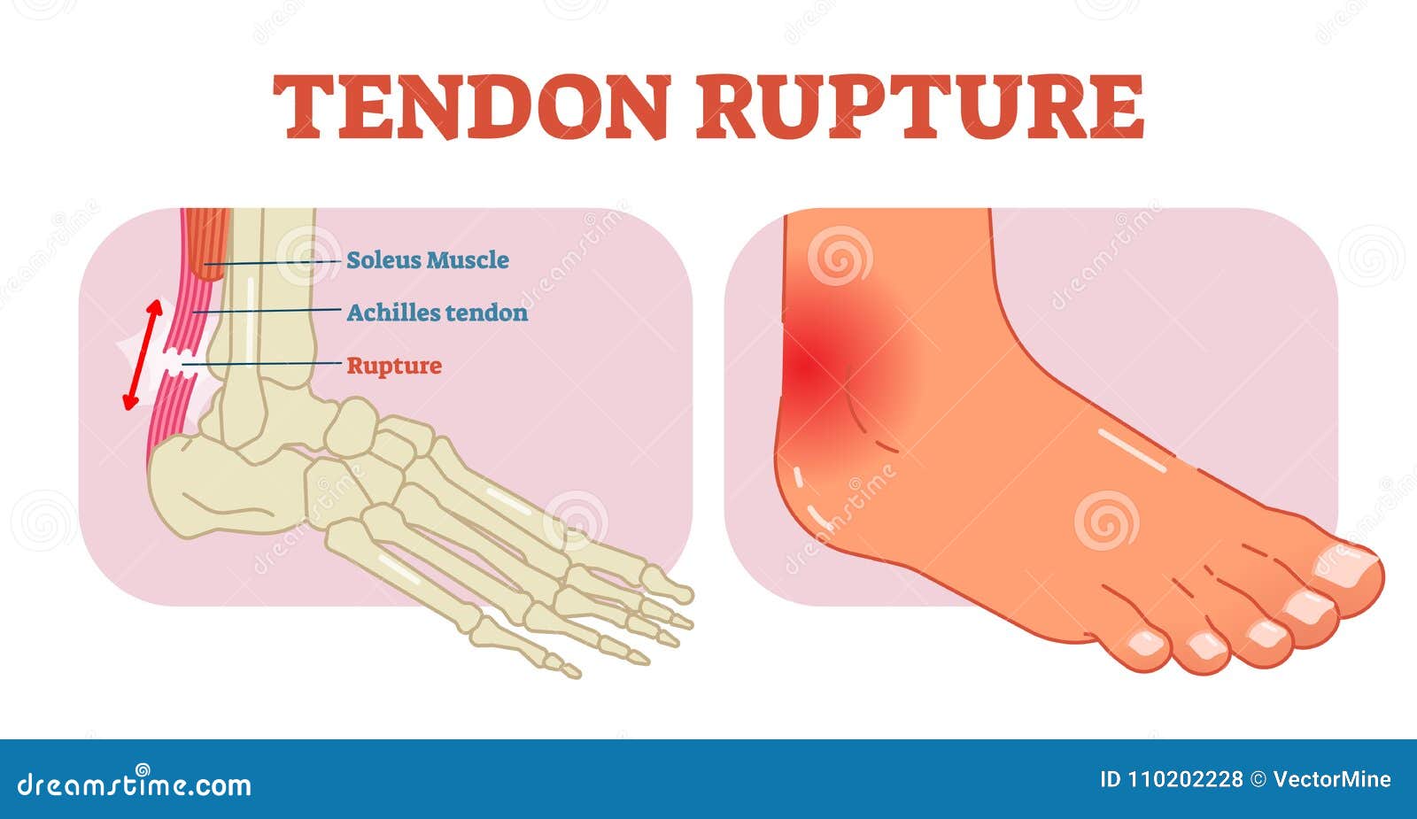
If you tear the biceps tendon at the shoulder, you may lose some strength in your arm and have pain when you forcefully turn your arm from palm down to palm up. Tendons are remarkably strong, having one of the highest tensile strengths found among soft tissues. The achilles tendon is the largest. Foot and ankle musculoskeletal key : The achilles tendon enables us to walk, without it we would not be able to raise our heels of the ground.
Tendon Ligament Bone And Cartilage Anatomy Physiology And Adaptations To Exercise And Training Veterian Key from veteriankey.com Tendons, located at each end of a muscle, attach muscle to bone. The achilles tendon is a tough band of fibrous tissue that connects the calf muscles to the heel bone (calcaneus). The achilles tendon is the strongest and largest tendon in the body. Ligaments and tendons are adapted in response to changes in mechanical stiffness. Foot anatomy diagram, foot joint diagram, foot sprain diagram, foot tendons and ligaments pain, leg tendon diagram, peroneal tendonitis, foot, foot anatomy diagram, foot joint diagram, foot sprain diagram, foot tendons and ligaments pain, leg tendon diagram, peroneal tendonitis. Fall on one point of shoulder and can rupture these ligaments with dislocation of ac joint. Your biceps tendons attach the biceps muscle to bones in the shoulder and in the elbow. Superficial posterior muscles of the forearm posterior compartment muscles of the forearm.
This sudden, tight, intense lower leg pain is sometimes called a charley horse.
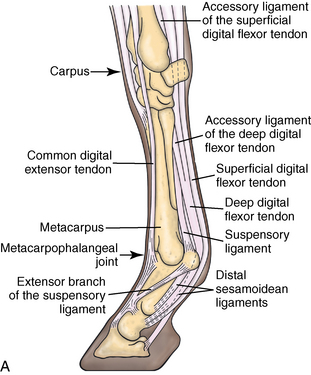
Foot anatomy diagram, foot joint diagram, foot sprain diagram, foot tendons and ligaments pain, leg tendon diagram. Your biceps tendons attach the biceps muscle to bones in the shoulder and in the elbow. The hip itself is a ball and socket joint, much like the shoulder.the structures necessary to create this joint are the socket, the joint capsule, muscle, ligaments, and the neck. A muscle's origin is where a tendon attaches it to the *less* movable bone. Jul 05, 2018 · the foot diagram has a complex structure made up of bones, ligaments, muscles, and tendons. Tendon diagrams and design force vectors. Tendons are the connection between bones and muscles. On the other hand, the insertion is where a tendon attaches that muscle to the *more* movable bone. When the muscles tighten (contract) arguably, the most important tendon is the achilles tendon, which allows the calf muscles to move. Foot anatomy diagram, foot joint diagram, foot sprain diagram, foot tendons and ligaments pain, leg tendon diagram, peroneal tendonitis, foot, foot anatomy diagram, foot joint diagram, foot sprain diagram, foot tendons and ligaments pain, leg tendon diagram, peroneal tendonitis. Possibly the most important tendon in terms of mobility is the achilles tendon. Allows the action of raising the foot. The achilles tendon is the largest.
Brings leg back to and across body. The hand incorporates countless muscles, bones, tendons and ligaments into simple motion and this chart covers them all. Superficial posterior muscles of the forearm posterior compartment muscles of the forearm. You can see a diagram of the achilles tendon below. Allows the foot to be turned inward and also supports the arch of the foot.
Tendon Rupture Anatomical Example Vector Illustration Diagram Educational Medical Scheme Stock Vector Illustration Of Physical Painful 110202228 from thumbs.dreamstime.com The bones of the hip include the femur, the ilium, the ischium, and the pubis. Intermediate back muscles and c. Browse 318 hand anatomy tendons stock photos and images available, or start a new search to explore more stock photos and images. The knee joint is a complex structure that involves bones. In the back and elsewhere in the body, tendons attach muscles to bones. Tendons are remarkably strong, having one of the highest tensile strengths found among soft tissues. Tendons are thick bands of tissue that connect muscles to bones. It is controlled by the obturator nerve.
Ligaments join the knee bones and provide stability to the knee:
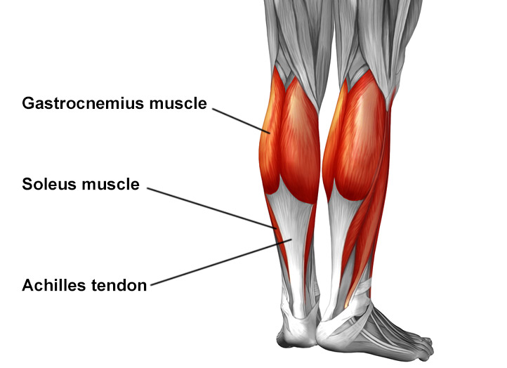
This important tendon in the back of the calf and ankle connects the plantaris, gastrocnemius, and soleus muscles to. Superficial posterior muscles of the forearm posterior compartment muscles of the forearm. For images of the muscle, click on each link under location. Diagram of tendons in forearm. 17 photos of the diagram of shoulder muscles and tendons. If you tear the biceps tendon at the shoulder, you may lose some strength in your arm and have pain when you forcefully turn your arm from palm down to palm up. Tendon, tissue that attaches a muscle to other body parts, usually bones.tendons are the connective tissues that transmit the mechanical force of muscle contraction to the bones; Intermediate back muscles and c. You can see a diagram of the achilles tendon below. The achilles tendon enables us to walk, without it we would not be able to raise our heels of the ground. Tendons attach muscles to bones. Allows the action of raising the foot. Possibly the most important tendon in terms of mobility is the achilles tendon.
Tendon, tissue that attaches a muscle to other body parts, usually bones.tendons are the connective tissues that transmit the mechanical force of muscle contraction to the bones; Tendon diagrams and design force vectors. Raises heal when leg is straight. Allows the foot to be turned inward and also supports the arch of the foot. One peroneal tendon attaches to the outer part of the midfoot, while the other tendon runs under the foot and attaches near the inside of the arch.
Achilles Tendon Pain Causes Diagnosis And Treatment Hss from www.hss.edu Tendons are remarkably strong, having one of the highest tensile strengths found among soft tissues. Tendons are thick bands of tissue that connect muscles to bones. The tendon travels along the inside of the forearm on the side of the small finger and crosses the wrist. Allows the action of raising the foot. Possibly the most important tendon in terms of mobility is the achilles tendon. You can see a diagram of the achilles tendon below. Tendon, tissue that attaches a muscle to other body parts, usually bones.tendons are the connective tissues that transmit the mechanical force of muscle contraction to the bones; A tendon is a band of tissue that connects a muscle to a bone.
The tendon runs down the back of your lower leg from the back of the knee to the heel.
The pubis, ischium, and ilium together constitute the pelvis while the thigh bone is the femur. The changes in ligaments and tendons generally occur more slowly than adaptation in bone, because ligaments and tendons have less vascular supply. Tendon, tissue that attaches a muscle to other body parts, usually bones.tendons are the connective tissues that transmit the mechanical force of muscle contraction to the bones; It attaches to the wrist bone, the pisiform, and as well as the 5th hand bone. Diagram of tendons in forearm. Jul 05, 2018 · the foot diagram has a complex structure made up of bones, ligaments, muscles, and tendons. The coracobrachialis muscle lies deep to the biceps brachii in the arm. Tendon diagrams and design force vectors. You can see a diagram of the achilles tendon below. The fcu tendon is one of two tendons that bend the wrist. The bones together make up the hip. Tendons, located at each end of a muscle, attach muscle to bone. For images of the muscle, click on each link under location.